Animal DevelopmentPage
4
4
The frog blastula is many cell layers thick. Cells of the dorsal lip originate in the gray crescent and invaginate to create the archenteron.
Cells continue to move from the embryo surface into the embryo by involution. These cells become the endoderm and mesoderm.
The blastopore encircles a yolk plug when gastrulation is completed.
The surface of the embryo is now ectoderm, the innermost layer is endoderm, and the middle layer is mesoderm.
Slide 23
Gastrulation in a frog embryo
Future ectoderm
Key
Future endoderm
Future mesoderm
SURFACE VIEW
Animal pole
Vegetal pole
Early gastrula
Blastopore
Blastocoel
Dorsal lip of blasto- pore
CROSS SECTION
Dorsal lip of blastopore
Late gastrula
Blastocoel shrinking
Archenteron
Blastocoel remnant
Archenteron
Blastopore
Blastopore
Yolk plug
Ectoderm
Mesoderm
Endoderm
Slide 24
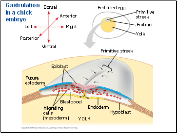
Gastrulation in the chick
The embryo forms from a blastoderm and sits on top of a large yolk mass.
During gastrulation, the upper layer of the blastoderm (epiblast) moves toward the midline of the blastoderm and then into the embryo toward the yolk.
The midline thickens and is called the primitive streak.
The movement of different epiblast cells gives rise to the endoderm, mesoderm, and ectoderm.
Slide 25
Gastrulation in a chick embryo
Endoderm
Future ectoderm
Migrating cells (mesoderm)
Hypoblast
Dorsal
Fertilized egg
Blastocoel
YOLK
Anterior
Right
Ventral
Posterior
Left
Epiblast
Primitive streak
Embryo
Yolk
Primitive streak
Slide 26
Organogenesis
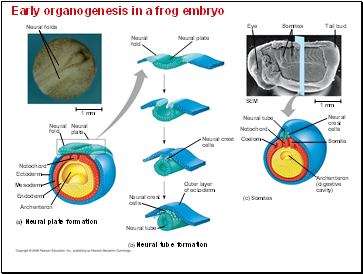
During organogenesis, various regions of the germ layers develop into rudimentary organs.
The frog is used as a model for organogenesis.
Early in vertebrate organogenesis, the notochord forms from mesoderm, and the neural plate forms from ectoderm.
Slide 27
Early organogenesis in a frog embryo
Neural folds
Tail bud
Neural tube
(b) Neural tube formation
Neural fold
Neural plate
Neural fold
Neural plate
Neural crest cells
Neural crest cells
Outer layer of ectoderm
Mesoderm
Notochord
Archenteron
Ectoderm
Endoderm
(a) Neural plate formation
(c) Somites
Neural tube
Coelom
Notochord
1 mm
1 mm
SEM
Somite
Neural crest cells
Archenteron (digestive cavity)
Somites
Eye
Slide 28
Contents
- A Body-Building Plan
- The Cortical Reaction
- Activation of the Egg
- Fertilization in Mammals
- Cleavage = Rapid Mitosis / No Mass change
- Cleavage in an echinoderm embryo
- Gastrulation
- Gastrulation in the frog
- Gastrulation in the chick
- Organogenesis
- Developmental Adaptations of Amniotes
- Amniote ExtraEmbryonic Membranes
- Mammalian Development
- The Cytoskeleton, Cell Motility, and Convergent Extension
- Role of Cell Adhesion Molecules and the Extracellular Matrix
- The developmental fate of cells depends on their history and on inductive signals
- Two general principles underlie differentiation
- Formation of the Vertebrate Limb
- You should now be able to
Last added presentations
- Madame Marie Curie
- Gravitation
- Mechanics Lecture
- History of Modern Astronomy
- Understanding Heat Transfer, Conduction, Convection and Radiation
- Heat-Energy on the Move
- Ch 9 Nuclear Radiation


