The Structure and Function of Large Biological MoleculesPage
9
9
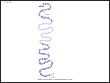
Fig. 5-21b
Amino acid
subunits
+H3N
Amino end
Carboxyl end
125
120
115
110
105
100
95
90
85
80
75
20
25
15
10
5
1
Slide 79
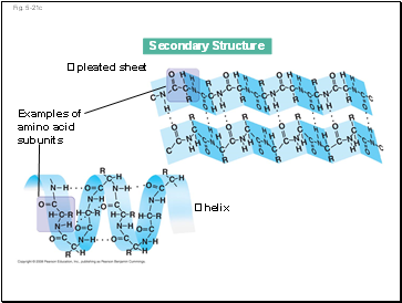
The coils and folds of secondary structure result from hydrogen bonds between repeating constituents of the polypeptide backbone
Typical secondary structures are a coil called an helix and a folded structure called a pleated sheet
Animation: Secondary Protein Structure
Slide 80
Fig. 5-21c
Secondary Structure
pleated sheet
Examples of
amino acid
subunits
helix
Slide 81
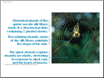
Fig. 5-21d
Abdominal glands of the
spider secrete silk fibers
made of a structural protein
containing pleated sheets.
The radiating strands, made
of dry silk fibers, maintain
the shape of the web.
The spiral strands (capture
strands) are elastic, stretching
in response to wind, rain,
and the touch of insects.
Slide 82
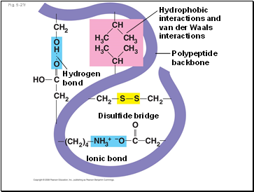
Tertiary structure is determined by interactions between R groups, rather than interactions between backbone constituents
These interactions between R groups include hydrogen bonds, ionic bonds, hydrophobic interactions, and van der Waals interactions
Strong covalent bonds called disulfide bridges may reinforce the protein’s structure
Animation: Tertiary Protein Structure
Slide 83
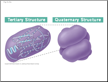
Fig. 5-21e
Tertiary Structure
Quaternary Structure
Slide 84
Fig. 5-21f
Polypeptide
backbone
Hydrophobic
interactions and
van der Waals
interactions
Disulfide bridge
Ionic bond
Hydrogen
bond
Slide 85
Fig. 5-21g
Polypeptide
chain
Chains
Heme
Iron
Chains
Collagen
Hemoglobin
Slide 86
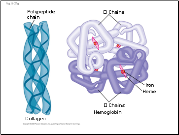
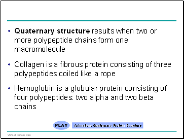
Quaternary structure results when two or more polypeptide chains form one macromolecule
Collagen is a fibrous protein consisting of three polypeptides coiled like a rope
Hemoglobin is a globular protein consisting of four polypeptides: two alpha and two beta chains
Animation: Quaternary Protein Structure
Slide 87
Contents
- The Molecules of Life
- The Synthesis and Breakdown of Polymers
- The Diversity of Polymers
- Sugars
- Polysaccharides
- Fats
- Phospholipids
- Steroids
- Polypeptides
- Protein Structure and Function
- The Roles of Nucleic Acids
- The Structure of Nucleic Acids
- The DNA Double Helix
- DNA and Proteins as Tape Measures of Evolution
- The Theme of Emergent Properties in the Chemistry of Life: A Review
Last added presentations
- Newton’s Laws of Motion
- Newton’s laws of motion
- Gravitation
- Waves & Sound
- Upcoming Classes
- Newton's laws of motion
- Sound

